Somatostatin Receptor Analogue Scintigraphy
Somatostatin receptor scintigraphy is not specific for carcinoids. Uptake occurs in other lesions with a high density of somatostatin receptors these include gastrinomas, glucagonomas, somatostatinomas, vasoactive intestinal polypeptide tumors, neural crest tumors , oat cell lung carcinomas, and lymphoproliferative disease . In addition, the possibility of uptake in areas of lymphocyte concentration in inflammatory states must be kept in mind. Approximately 20% of gastrinomas are missed during somatostatin receptor analogue scintigraphy.
Somatostatin receptor analogue scintigraphy has had the greatest impact on the diagnosis of gastrinomas it has a sensitivity and a specificity of 80-90% for the detection of both primary and metastatic sites. Somatostatin receptor analogue scintigraphy may depict subcentimeter liver metastases with a high signal-to-noise ratio. Somatostatin receptor analogue scintigraphy has been reported to show uptake in insulinomas, glucagonomas, small-cell lung cancer, thyroid cancer, and carcinoids.
Somatostatin receptor analogue scintigraphy may prove useful in the treatment of patients with hypergastrinemic states who have increased incidence of gastric carcinoids. In patients with multiple endocrine neoplasia type 1 , localization in the upper abdomen may not be associated with a pancreatic endocrine tumor instead, it may be caused by a gastric carcinoid.
Differentiation From Focal Fat
Results of sulfur colloid scans are usually normal, with focal fatty infiltration and focal fatty sparing, because Kupffer cells are not affected by steatosis.
Xenon-133 is highly fat-soluble. After its inhalation, it characteristically concentrates in areas of focal fatty infiltration. The intensity of the radionuclide concentration is related to the fat content.
Treatment For Metastatic Liver Cancer
Specific treatment for metastatic liver cancer will be determined by your doctor based on:
- Your age, overall health, and medical history
- Extent of the disease
- Your tolerance of specific medicines, procedures, or therapies
- Expectations for the course of the disease
- Your opinion or preference
Treatment may include:
- Surgery. In some cases, surgery may be used to remove cancerous tissue from the liver. However, the tumor must be small and confined.
- Radiation therapy. Radiation therapy uses high-energy rays to kill or shrink cancer cells.
- Chemotherapy. Chemotherapy uses anticancer drugs to kill cancer cells.
Don’t Miss: Antiviral Drugs For Hepatitis A
Questions For Your Doctor
When you’re diagnosed with any kind of cancer, you’re bound to have a lot of questions for your doctor, such as:
- What treatments will work best for me? Whatâs involved?
- For how long will I need treatment?
- What’s my outlook?
- Should I consider a clinical trial?
- Should I get a second opinion? Will you recommend someone?
- How often should I see you for follow-ups?
Intraoperative Us And Laparoscopic Us

IOUS is an important diagnostic tool in patients undergoing hepatic resection for colorectal metastases. IOUS allows careful evaluation of the normal liver segments to exclude occult metastases in the segments that will be left in situ. The high accuracy of IOUS is a result of the contact scanning possible with a high-frequency transducer and color-flow Doppler imaging with this technique, the complete organ may be covered without artifact. IOUS depicts 25-35% more lesions than does preoperative US. Most significantly, 40% of the lesions detected by means of IOUS are neither visible nor palpable and would presumably have been missed with other means.
IOUS has also been shown to be a sensitive means of detecting HCC, particularly if US contrast agents are used to improve Doppler images. IOUS has been used as an aid to liver resection since the end of the 1970s. This approach has been particularly useful in the resection of tumors from a cirrhotic liver in such cases, conventional resection methods would result in high mortality and morbidity rates. IOUS combines the needs for adequate tumor resection with sparing of the liver parenchyma.
IOUS also permits an accurate 3-dimensional reconstruction of the relationships between the tumor, the hepatic veins, and the portal branches. Moreover, the portal venous branches are used as landmarks in defining the resection line. This finding is fundamental for planning the surgical strategy.
Also Check: What Is Included In A Hepatitis Panel
Tc Sulfur Colloid Scintigraphy
Sulfur colloid scintigraphy has largely been abandoned as an imaging test for liver metastases despite its reasonably high sensitivity for the detection of metastatic disease . The lesions appear as photon-deficient defects, which are nonspecific. Also, the sensitivity of planar imaging decreases dramatically for surface lesions smaller than 2 cm and deeper lesions smaller than 3-4 cm in diameter. A genuine liver lesion within a fatty liver may be missed or mischaracterized on other imaging studies. In such instances, sulfur colloid scanning may be useful in confirming or excluding a mass lesion in the liver.
Sulfur colloid scans may still be indicated when the CT findings are nondiagnostic because of a fatty liver. SPECT imaging has improved the overall sensitivity but at the expense of reduced specificity because of problems with distinguishing small lesions near the heart or intrahepatic vessels. Because the Kupffer cells are unaffected by fatty infiltration, sulfur colloid scans are typically normal. Sulfur colloid scintigraphy is highly sensitive and specific for focal fatty infiltration.
Hepatocellular neoplasms such as a hepatocellular adenoma and hepatocellular carcinoma may also have Kupffer cells, and they may demonstrate sulfur colloid uptake. Typically, hepatic adenomas appear photopenic on sulfur colloid scans, but colloid uptake in a liver lesion does not exclude a hepatic adenoma.
Sensitivity And Specificity Of Mri
Technically, MRI is as sensitive as CT in the detection of liver metastases. The use of ultrafast techniques has certainly increased the sensitivity of MRI, although it is still inferior to CTAP. In a number of settings, MRI is superior to other imaging techniques. Hemangiomas are reliably diagnosed with MRI, and more importantly, they are more easily differentiated from metastases with MRI than with other imaging modalities.
MRI is said to be the best modality in the diagnosis of focal nodular hyperplasia it has a sensitivity of 70% and a specificity of 98%. The central scar is more often detected by MRI than by CT. One limiting factor of gadolinium-enhanced MRIs of the liver is that the liver must be imaged repetitively with T1-weighted gradient-echo sequences during hepatic arterial, portal venous, and delayed phase of contrast enhancement.
Gadolinium-enhanced study is always performed in the phase that shows the greatest differences in the distribution of contrast agent between normal tissues and abnormal tissues. For all practical purposes, this means such studies are performed during the portal venous phase. Therefore, a time limit exists during which imaging may be performed with gadolinium-based and other extracellular contrast agents. The time-limiting factor may be overcome through the use of tissue-specific contrast agents.
You May Like: Symptoms Of Advanced Hepatitis C
What Is Metastatic Liver Cancer
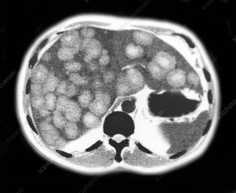
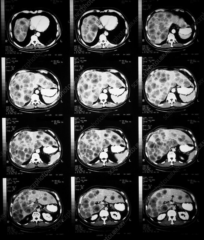
Metastatic liver cancer, also known as secondary liver cancer, begins as a primary cancer in another organ outside of the liver, and eventually migrates to the liver.
In fact, the liver is the most common site for cancers to spread. Most of these originate from cancers of the eye, colon, rectum, pancreas, stomach, esophagus, breast, lung, melanoma and some other less common sites.
Bull’s Eye Or Target Metastases
In bull’s eye, or target, metastases, the halo is most probably related to a combination of compressed normal hepatic parenchyma around the mass and a zone of cancer cell proliferation. The presence of a halo usually suggests aggressive behavior. Bronchogenic carcinoma characteristically causes target-type metastases. However, this pattern is nonspecific and may be found with metastases from the breast and colon, as well as primary malignant liver neoplasms and benign liver neoplasms . A similar appearance has been described with liver abscesses.
Recommended Reading: What Does It Mean If You Have Hepatitis C Antibodies
How Is Liver Hepatoma Diagnosed
In addition to a complete medical history and physical examination, diagnostic procedures for a liver hepatoma may include the following:
-
Liver function tests. A series of special blood tests that can determine if the liver is functioning properly.
-
Abdominal ultrasound . A diagnostic imaging technique that uses high-frequency sound waves to create an image of the internal organs. Ultrasounds are used to view internal organs of the abdomen, such as the liver, spleen, and kidneys and to assess blood flow through various vessels.
-
Computed tomography scan . A diagnostic imaging procedure using a combination of X-rays and computer technology to produce horizontal, or axial, images of the body. A CT scan shows detailed images of any part of the body, including the bones, muscles, fat, and organs. CT scans are more detailed than general X-rays.
-
Hepatic angiography. X-rays taken after a substance in injected into the hepatic arteries.
-
Liver biopsy. A procedure in which tissue samples from the liver are removed from the body for examination under a microscope.
Symptoms Of Metastases To The Liver
Often, secondary liver cancer does not cause symptoms during early stages. However, as the disease progresses, symptoms associated with liver disease often develop, including:
- Pain in the upper abdomen on the right side the pain may extend to the back and shoulder
- Swollen abdomen
- Weight loss, loss of appetite and/or feelings of fullness
- Weakness and/or fatigue
Don’t Miss: How Does Hepatitis Affect The Body
Treatment Of Lymphatic Metastasis
Treatment of lymphatic metastasis, given its close relation with the growth of the primary tumor, is the same as for the original lesion: primary resection with exeresis of the nodes of the lymphatic area that drain the tumor. If there is a tumor recurrence in the nodes, the patient is classified as stage IV, which means that there will be the corresponding treatment: chemotherapy and surgery and radiotherapy, which will be provisionally palliative.
Survival And Life Expectancy

Liver metastases usually originate in colorectal cancer, which has a worldwide incidence of 1,400,000 new cases per year. Of these, between 50 and 70% will present liver metastases at some point in their disease and are usually the cause of a worse disease progression.
Up until a few years back, patients with liver metastases could hardly be treated, as the chances of cure or increased survival were slim.
However, currently, resection of metastases is the most important prognostic factor for patient survival, according to numerous studies. The goal of treatmentis to achieve complete resection of liver metastases, emerging as the main treatment option.
Advances in surgical techniques have been key to the treatment of liver metastases. These new surgical techniques such as complete resection have made it possible to increase the life expectancy of these patients, achieving prolonged survival. According to studies, there is a 70% 5-year survival in patients with complete resection.
Furthermore, newer chemotherapy techniques allow reduction of lesions and complete resection of lesions that were previously unresectable. This new approach has been shown to achieve up to 87% complete resection after chemotherapy treatment.
On the other hand, new imaging techniques and evaluation of liver function have also made it possible to define new strategies for the management of metastases that have shown to increase the life expectancy of patients.
You May Like: How Do You Get Hepatitis A B C
Liver Resections Per Pathology Laboratory
The number of pathology laboratories examining liver resection specimens decreased from 37 in 2001 to 31 in 2010. Especially the low volume’ and sporadic centers’ decreased from 15 respectively 11 in 2001, to 10 respectively 2 in 2010 . A median of 28 resection specimens were evaluated annually in high volume centers’. Middle volume centers’ examined 13 resection specimens per year, and in low volume centers’, 4 resection specimens were evaluated yearly.
Fig. 1
Amount of liver resections performed in high , middle and low volume centers . Below the figure is the number of pathology laboratories involved in examining liver resection specimens per year.
In high volume centers’, resection specimens with multiple metastases and non-CRLM were more often examined than in middle’ and low volume centers’. Furthermore, in high volume centers’ patients were younger at the time of liver resection compared to low’ and middle volume centers’ . No differences were observed in the amount of complete resections and in the size of the liver metastases between the high’, middle’ and low volume centers’ .
Table 2
Patterns in resection characteristics in low , middle and high volume centers
How Are Liver Metastases Treated
Secondary liver cancer is generally treated based on the primary cancer, taking into consideration what care the patient has already received. Cases in which only one or a few areas of cancer are found, surgical removal may be an option. Other treatment methods work toward slowing the growth and managing the problems caused by liver metastases. Chemotherapy is the most common treatment approach. In some cases, it may even shrink the cancer enough to be surgically removed. Other treatment methods include: targeted therapy, ablation therapy, radiation therapy, and hormonal therapy.
You May Like: What Does Hepatitis C Mean
Does Treatment Cure Metastatic Cancer
In some situations, metastatic cancer can be cured, but most commonly, treatment does not cure the cancer. But doctors can treat it to slow its growth and reduce symptoms. It is possible to live for many months or years with certain types of cancer, even after the development of metastatic disease.
How well any treatment works depends on:
-
The type of cancer
-
How far the cancer has spread and where it is located
-
How much cancer there is
-
If the cancer is growing quickly or slowly
-
The specific treatment
-
How the cancer responds to treatment
It is important to ask your doctor about the goals of treatment. These goals may change during your care, depending on whether the cancer responds to the treatment. It is also important to know that pain, nausea, and other side effects can be managed with the help of your health care team. This is called palliative care and should be a part of any treatment plan.
Research shows that palliative care can improve the quality of your life and help you feel more satisfied with the treatment you receive. Learn more about palliative care, or supportive care.
What Are Noncancerous Liver Tumors
Noncancerous tumors are quite common and usually do not produce symptoms. Often, they are not diagnosed until an ultrasound, computed tomography scan, or magnetic resonance imaging scan is performed. There are several types of benign liver tumors, including the following:
-
Hepatocellular adenoma. This benign tumor is linked to the use of certain drugs. Most of these tumors remain undetected. Sometimes, an adenoma will rupture and bleed into the abdominal cavity, requiring surgery. Adenomas rarely become cancer.
-
Hemangioma. This type of benign tumor is a mass of abnormal blood vessels. Treatment is usually not required. Sometimes, infants with large liver hemangiomas require surgery to prevent clotting and heart failure.
Don’t Miss: Hepatitis B How Long Does It Last
What Is Metastatic Cancer
In metastasis, cancer cells break away from where they first formed , travel through the blood or lymph system, and form new tumors in other parts of the body. The metastatic tumor is the same type of cancer as the primary tumor.
Cancer that spreads from where it started to a distant part of the body is called metastatic cancer. For many types of cancer, it is also called stage IV cancer. The process by which cancer cells spread to other parts of the body is called metastasis.
When observed under a microscope and tested in other ways, metastatic cancer cells have features like that of the primary cancer and not like the cells in the place where the metastatic cancer is found. This is how doctors can tell that it is cancer that has spread from another part of the body.
Metastatic cancer has the same name as the primary cancer. For example, breast cancer that spreads to the lung is called metastatic breast cancer, not lung cancer. It is treated as stage IV breast cancer, not as lung cancer.
Sometimes when people are diagnosed with metastatic cancer, doctors cannot tell where it started. This type of cancer is called cancer of unknown primary origin, or CUP. See the Carcinoma of Unknown Primary page for more information.
How Does Liver Cancer Spread
Abnormal cells usually die off and are replaced by healthy cells. Sometimes, instead of dying off, these cells reproduce. As the cell numbers grow, tumors begin to form.
The overgrowth of abnormal cells can invade nearby tissue. By traveling through lymph or blood vessels, the cancerous cells can move all around the body. If they invade other tissues or organs, new tumors can form.
If the cancer invades nearby tissue or organs, its considered regional spread. This can happen during stage 3C or stage 4A liver cancer.
In Stage 3C, a liver tumor is growing into another organ . A tumor could also be pushing into the outer layer of the liver.
In Stage 4A, there are one or more tumors of any size in the liver. Some have reached blood vessels or nearby organs. Cancer is also found in nearby lymph nodes.
Cancer that has metastasized to a distant organ, such as to the colon or lungs, is considered stage 4B.
In addition to telling how far the cancer has spread, staging helps determine which treatments may be most helpful.
Remission means that you have fewer or no signs or symptoms of liver cancer after treatment. It doesnt mean that youre cured. You still might have cancer cells in your body, but your disease is under control.
Thanks to new targeted therapies like sorafenib , a very small percentage of people with late-stage liver cancer may go into complete remission.
Recommended Reading: Can You Get Hepatitis C From Alcohol
How Do Doctors Diagnose Metastasis
If you already had cancer treatment for non-metastatic cancer, you probably have a follow-up care plan. You will see your doctor for regular checkups. Specific tests may be done to look for metastases.
Alternatively, some people already have metastases when they are first diagnosed with cancer. In this situation, the metastases are usually found during the initial tests to stage the cancer.
Cancer may cause symptoms such as pain or shortness of breath. Sometimes these symptoms will lead your doctor to do necessary tests to find the metastases.